41 mushroom diagram labeled
Start studying Parts of a mushroom. Learn vocabulary, terms, and more with flashcards, games, and other study tools. Try our Mushrooms Life Cycle Worksheets! You'll be amazed at what your child can retain by using simple diagrams and label charts to get their curiosity jumping! Building key science skills is important at an early age — it keeps them curious while strengthening their retention and memory with fascinating facts.
Find a mushroom online or in real life, then draw and label the parts. Knowing these helps to identify different species. Challenge Yourself. Be Awesome. Join for Free. See All Challenges. Challenge. Draw and label a mushroom +2 XP. Easy. Your Challenge.
Mushroom diagram labeled
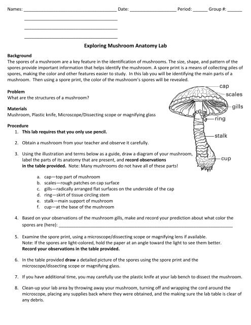
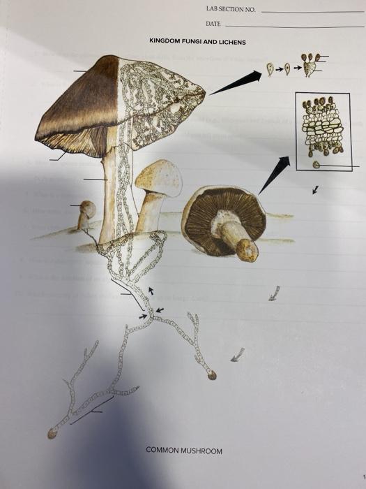
Draw a diagram of the mushroom in the lab book, labeling the cap, stem and gills. If the gills are not visible, remove the tissue (it's called a veil) protecting them gently with the forceps. Be careful not to touch the gills with the ... Draw and label the gills, basidia and spores in your lab book. Make sure to record the magnification. Mushroom Compound Light Microscope Forceps . Dissecting Microscope Microscope slide Cover slip. Water Eye Dropper Paper towels. Procedure: 1. Get your mushroom and place it on the paper towels in front of you. Examine it closely. On a sheet of paper draw a diagram of your mushroom, labeling the cap, stem and gills Mushroom anatomy labeled biology diagram vector illustration. 1. Editable Vector .AI file. 2. Editable Vector .EPS-10 file. 3. High-resolution JPG image. Use for everything except reselling item itself. Description: Mushroom anatomy labeled biology diagram vector illustration.
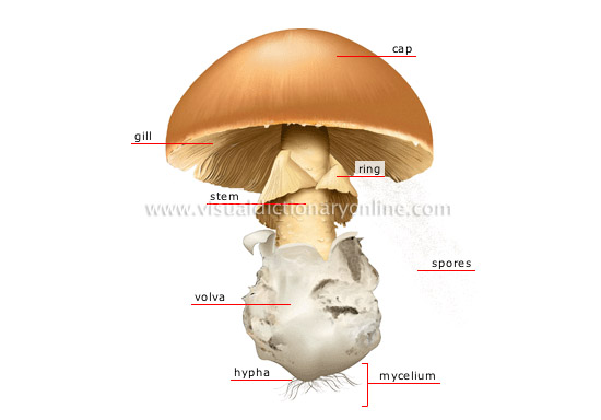
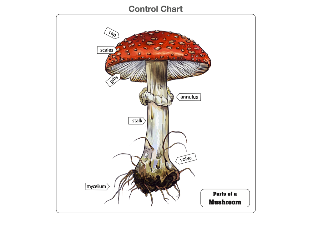
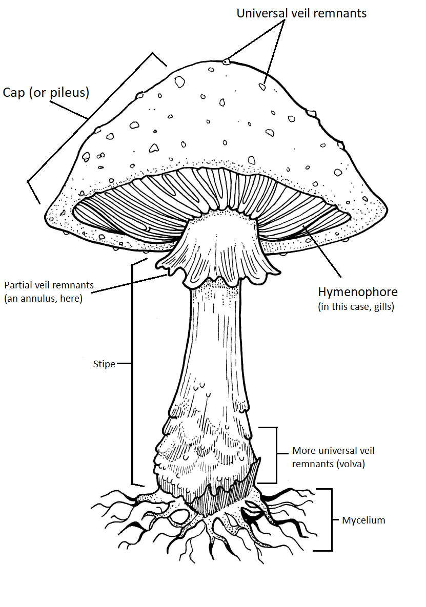
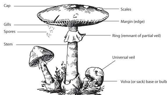
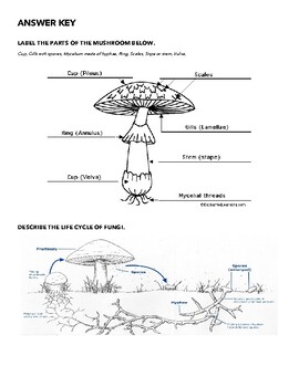
Mushroom diagram labeled. Mushroom anatomy labeled biology diagram vector illustration. Forest nature exploring and education. Spore bearing fruiting body of a fungus. Structural drawing scheme as learning information. Mushroom Anatomy Read the definitions then label the mushroom diagram, including the cap (pileus), cup (volva), gills (lamellae), mycelial threads, ring (annulus), scales, and stem (stape). Answers: Plant Anatomy Printout: Simple Label Me! Label the simple plant anatomy diagram using the glossary of plant terms. Words: leaf, flower, stem, root ... Typically, a mushroom has six different parts. These include: Cap: This is the part that gives the fungi its umbrella shape. The cap comes in a variety of colors, including white, brown, and yellow. In the same way that umbrellas protect us from the heat of the sun, rain, and other harsh weather conditions, the mushroom cap protects the pores ... Male Testicles Anatomy diagram. Male Testicles Anatomy diagram. In this image, you will find a Ligamentous remnant of processes vaginalis, Vas deferens, Head of the epididymis, Efferent ductules, Rete testis in mediastinum testis, Body of the epididymis, Tail of epididymis, Capsule, Tunica vaginalis, Seminiferous tubule, Straight tubule in it.
About this Quiz. This is an online quiz called Mushroom Diagram. There is a printable worksheet available for download here so you can take the quiz with pen and paper. This quiz has tags. Click on the tags below to find other quizzes on the same subject. mushroom. The cap of the mushroom is the topmost part. This is a thumbnail of the label the mushroom anatomy diagram. Label the cap stipe and gills. Cap pileus the top part of the mushroom. Rhizopus and mucor are the common saprotrophic fungi that attack a variety of food stuffs. Mucor pusillus causes infection of internal organs in human beings. This will also help you to draw the structure and diagram of the fungal cell. (a) The Cell Wall of the Fungal Cell: The composition of cell wall is variable among the different groups of fungi or between the different species of the same group. In the majority of fungi, the wall lacks cellulose but contains a form of chitin known as the fungus ... Find Mushroom Anatomy Labeled Biology Diagram Vector stock images in HD and millions of other royalty-free stock photos, illustrations and vectors in the Shutterstock collection. Thousands of new, high-quality pictures added every day.
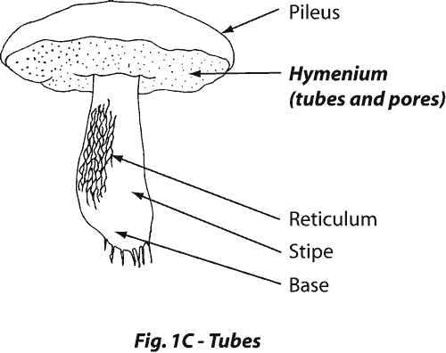
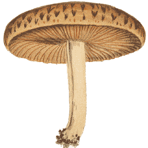
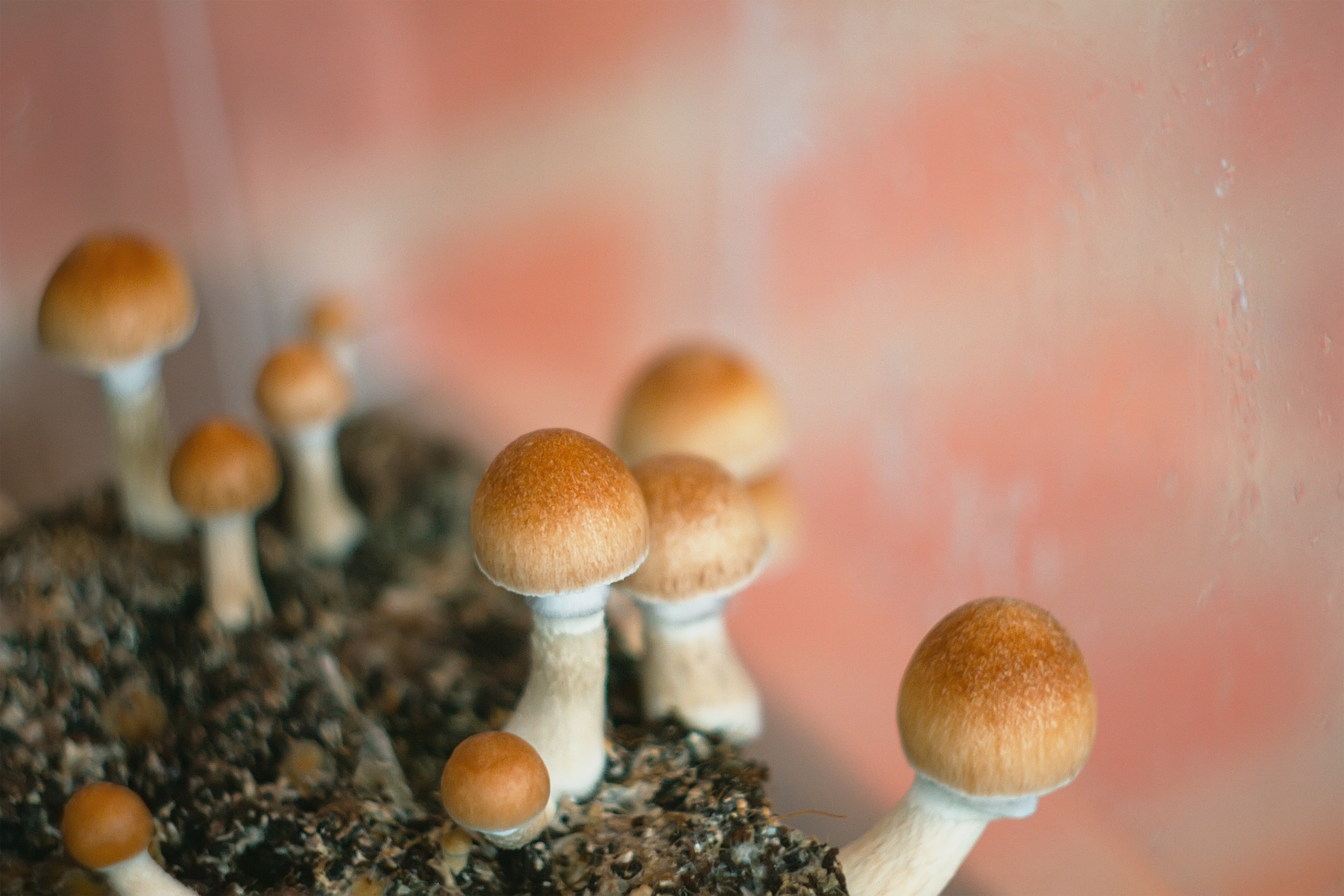
A. mellea (Honey Mushroom) is an edible mushroom which is a serious root parasite of both hardwoods and conifers. The fungus sends rhizomorphs into the phloem of the host and hence block the food supply. 8. Hallucinogens: Psilocybe mexicana (Sacred Mushroom) has hallucinating properties similar to LSD. Mushroom anatomy read the definitions then label the mushroom diagram including the cap pileus cup volva gills lamellae mycelial threads ring annulus scales and stem stape. Agaricus edible mushrooms puccinia rust fungi ustilago smut fungi polyporus bracket fungi candida etc. Gently wiggle andor twist the stipe until it breaks away from the cap. Parts of Mushrooms. Mushrooms have a variety of different parts. Mushroom's don't always look like the diagram below, but it's a good place to start when looking at what exactly a mushroom is and how it grows. Cap: The cap is the top of the mushroom (and often looks sort of like a small umbrella). Mushroom caps can come in a variety of colors ... Lisa Romerein/Photolibrary/Getty Images. The parts of the mushroom are the cap, gills or pores, spores, stem, ring, volva, mycelium and hypha. The mushroom can be divided into underground and aboveground sections. The cap of the mushroom is the topmost part. It can be conical, flat or spherical and have a variety of textures depending on the ...
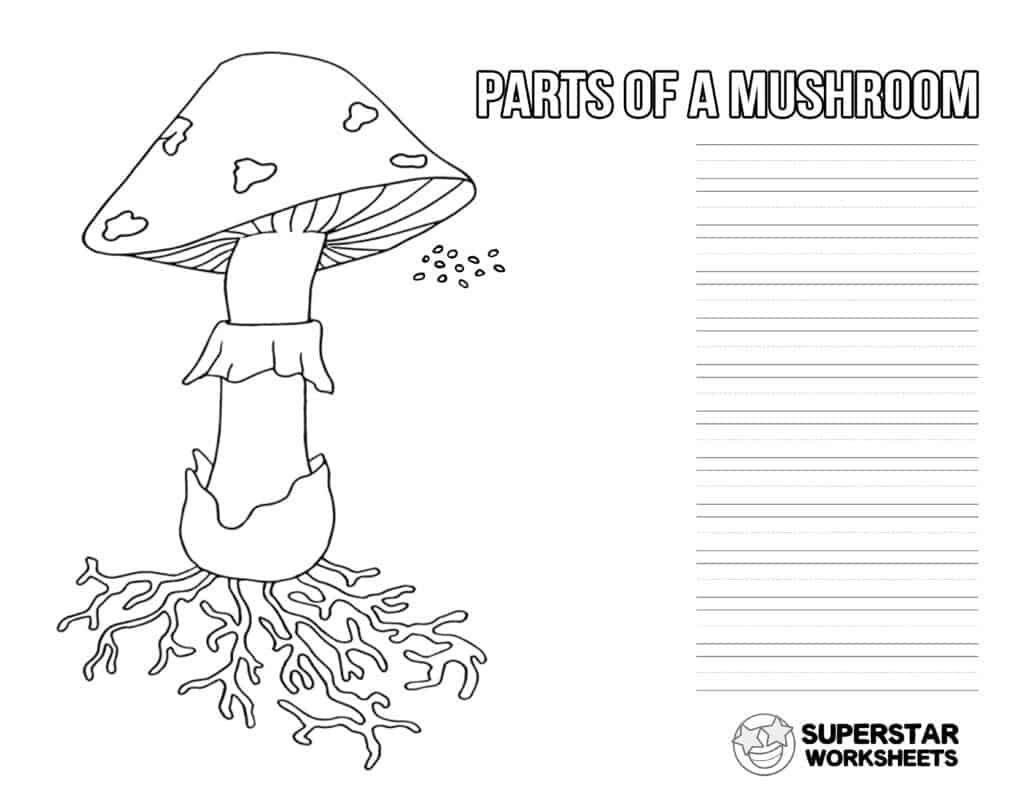
Mushroom Life Cycle Worksheets Superstar Worksheets
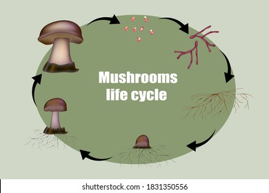
Mushroom anatomy labeled biology diagram vector illustration. Forest nature exploring and education. Spore bearing fruiting body of a fungus. Structural. Diagram mushroom anatomy life cycle stages. From spore release to inoculation, germination, mycelial expansion and hyphal knot to the primordia formation.
Wch Clinical Toxinology Resources
iStock Mushroom Anatomy Labeled Biology Diagram Vector Illustration Stock Illustration - Download Image Now Download this Mushroom Anatomy Labeled Biology Diagram Vector Illustration vector illustration now. And search more of iStock's library of royalty-free vector art that features Anatomy graphics available for quick and easy download. Product #: gm1204471308 $ 12.00 iStock In stock
1
Mushroom anatomy life cycle stages diagram, vector illustration labeled circular scheme. From spore release to inoculation, germination, mycelial expansion and hyphal knot to the primordia formation. 1 credit. Essentials collection. Everyday photos and illustrations, for just 1 credit. $12.
Exploring Mushroom Anatomy Lab 2009 2010
Read the definitions below, then label the mushroom diagram. This is a thumbnail of the Label the Mushroom Anatomy Diagram. The full-size printout is available only to site members. Cap (Pileus) - The top part of the mushroom. Cup (Volva) - A cup-shaped structure at the base of the mushroom. The ...
Plants Gardening Plants Mushroom Structure Of A Mushroom Image Visual Dictionary Online
The mushroom is composed of an underground part (mycelium) and an aboveground, often edible part that is also the reproductive organ. previous. next. spores Microscopic seeds acting as reproductive agents; they are usually released into the air and fall on a substrate to produce a new mushroom. stem Axis supporting the mushroom's cap. ...
Mycelium
Illustration of Mushroom anatomy labeled biology diagram vector illustration. Forest nature exploring and education. Spore bearing fruiting body of a fungus. Structural drawing scheme as learning information. vector art, clipart and stock vectors. Image 139640560.

Fungus Form And Function Of Fungi Britannica
Mushroom Nomenclature - Cards. Mushroom Nomenclature Cards are formatted in a 3-part card series with blackline master included. The 9 parts of the mushroom: mushroom, scales, volva, stalk, ring, pores, tubes, cap, and gills. 9 cards with labels 9 cards without labels 9 labels 1 blackline master Learn how to use Montessori nomenclature.

Question Video Labeling The Structures Of A Fungus Nagwa
A big thanks to all current and future patrons who are helping fund this science communication outreach via Patreon: http://bit.ly/2SfmkphThis is my re-relea...
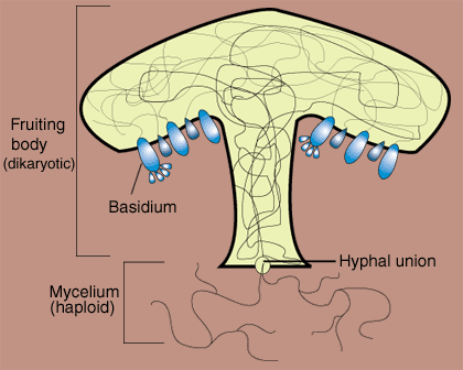
Fungi Basidiomycota The Club Fungi Sparknotes
1. Get your mushroom and place it on the paper towels in front of you. Examine it closely. On the bottom of this lab, draw a diagram of your mushroom, labeling the cap, stem and gills. If the gills are not visible, remove the tissue (it's called a veil) protecting them gently with your forceps. Be careful not to touch the gills with the forceps. 2.

Mushroom Kenya Mushroom And Its Parts Join Mushroom Kenya And Learn More Www Mushroomkenya Co Ke Facebook
On the back of this sheet, (a) draw a diagram of the mushroom you see. Label the cap, stipe, and gills. (see diagram in data section) 2.) Grasp the cap firmly with one hand and the stipe with the other. Gently wiggle and/or twist the stipe until it breaks away from the cap. 3.) Peel away some strips from the stipe (like string cheese).

Mushroom Anatomy Labeled Biology Diagram Vector Illustration Stock Vector Illustration Of School Info 171697068
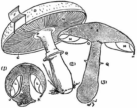
Mushroom Structure: Teacher's notes After learning about fungi in lessons; this exercise is a fun way for pupils ... (and at 8 o'clock under the label) still vertical. You can get the gills to re-orient themselves by putting the ... The diagrams below show the front face of this. Cap context (= cap flesh) Hymenial surface (covered with ...
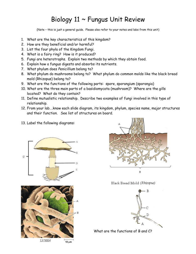
Biology 11 Fungus Unit Review
Mushroom anatomy labeled biology diagram vector illustration. 1. Editable Vector .AI file. 2. Editable Vector .EPS-10 file. 3. High-resolution JPG image. Use for everything except reselling item itself. Description: Mushroom anatomy labeled biology diagram vector illustration.

Mushroom Diagram Diagram Quizlet
Mushroom Compound Light Microscope Forceps . Dissecting Microscope Microscope slide Cover slip. Water Eye Dropper Paper towels. Procedure: 1. Get your mushroom and place it on the paper towels in front of you. Examine it closely. On a sheet of paper draw a diagram of your mushroom, labeling the cap, stem and gills
Solved Label This Diagram Of The Life Cycle Of A Mushroom Chegg Com
Draw a diagram of the mushroom in the lab book, labeling the cap, stem and gills. If the gills are not visible, remove the tissue (it's called a veil) protecting them gently with the forceps. Be careful not to touch the gills with the ... Draw and label the gills, basidia and spores in your lab book. Make sure to record the magnification.
Montessori Materials Parts Of A Mushroom Puzzle

Structure And Physiology Of Fungi
Mushroom Diagram Images Stock Photos Vectors Shutterstock

Mushroom Body Structure Mushroom Lifecycle Information About Mushrooms Mushrooms 101
1
Fs Usda Gov

Diagrammatic Representation Of Mushroom Life Cycle Download Scientific Diagram
Solved Label Fully The Accompanying Drawings Of A Mushroom Chegg Com
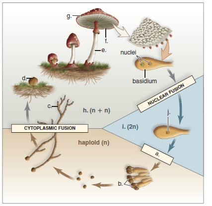
Solved Consider This Diagramof The Life Cycle Of A Mushroom Forming Basidiomycete Label The Haploid Hyphae Dikaryotic Hyphae Basidiocarp Basidi Course Hero

Fungi Structure Ck 12 Foundation
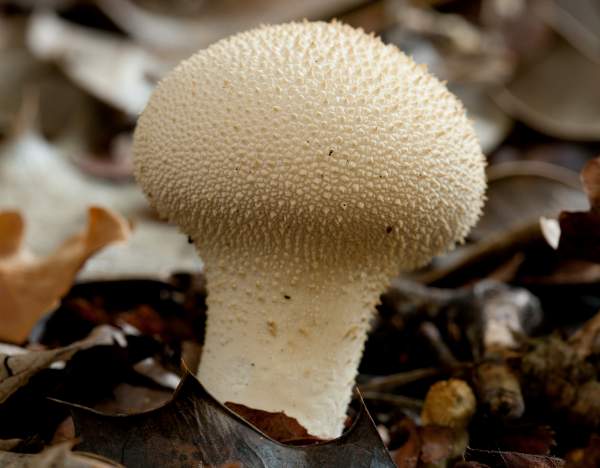
Lycoperdon Perlatum Common Puffball Identification
1

Diagram Of A Mushroom

Mushroom Body Structure Mushroom Lifecycle Information About Mushrooms Mushrooms 101

Wch Clinical Toxinology Resources

Mushroom Anatomy Understanding Caps Stems R R Cultivation
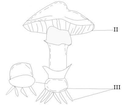
The Part Labeled Iii In The Above Diagram Is Erudites Academy
Mushroom Labeled Diagram Anatomy And Structure
Agaricus Bisporus The Commercial Mushroom Inanimate Life
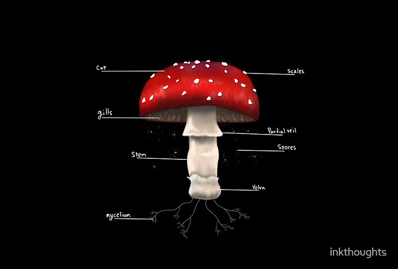
Labeled Mushroom By Inkthoughts Redbubble

Clip Art Vector Vector Diagram Showing Parts Of Mushroom Whole Plant Agricultural Infographic Amanita Muscaria Scheme With Labels For Education Of Biology Stock Eps Gg101847955 Gograph

Parts Of Mushrooms
1
What Are Magic Mushrooms Psychedelic Science Review
Mushroom Classification Using Machine Learning By Shravan Adulapuram Analytics Vidhya Medium
Mushroom And Fungi Life Cycle Diagram Label And Describe Tpt

Mushroom Anatomy Labeled Biology Diagram Vector Illustration Stock Illustration Download Image Now Istock


Comments
Post a Comment