42 neuromuscular junction diagram labeled
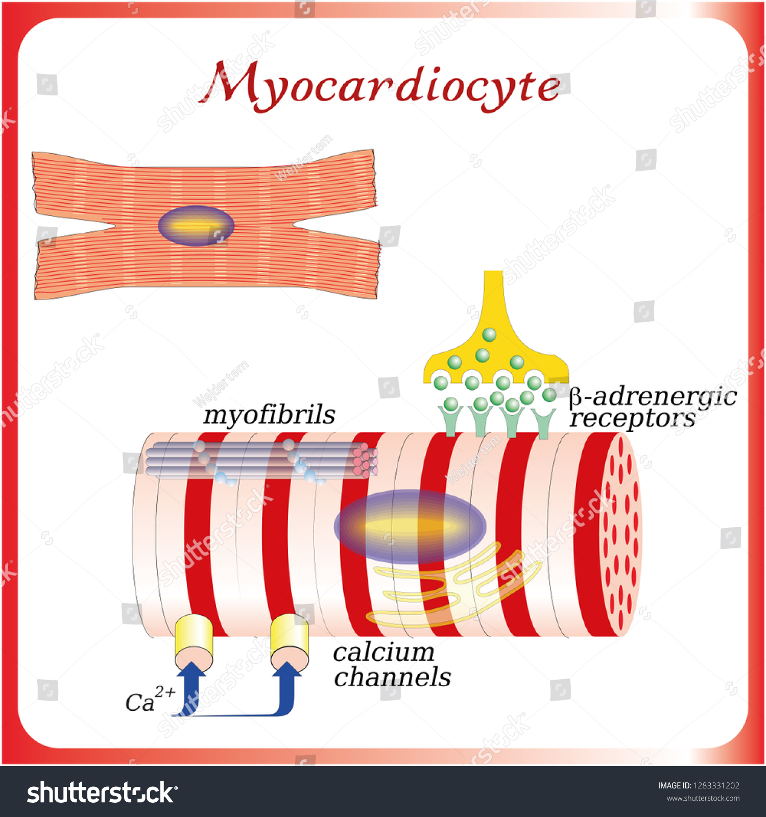
Art Print of Neuromuscular junction vector illustration scheme. Labeled medical infographic. Motor neuron and muscle cell structure closeup. Diagram with myofibril and muscle fibers. | bwc60977910. NEUROMUSCULAR JUNCTION LABELING (6 POINTS) Label the axon, motor end plate (neurotransmitter receptor), calcium channel, synaptic vesicles, neurotransmitter, and synapse in the diagram below. 1. Neurotransmitter 2. synapse 3. Motor end plate 4. Calcium channel 5. Synaptic vesicles 6. Axon Answer these questions: 1.
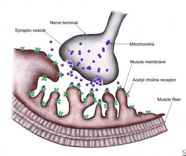
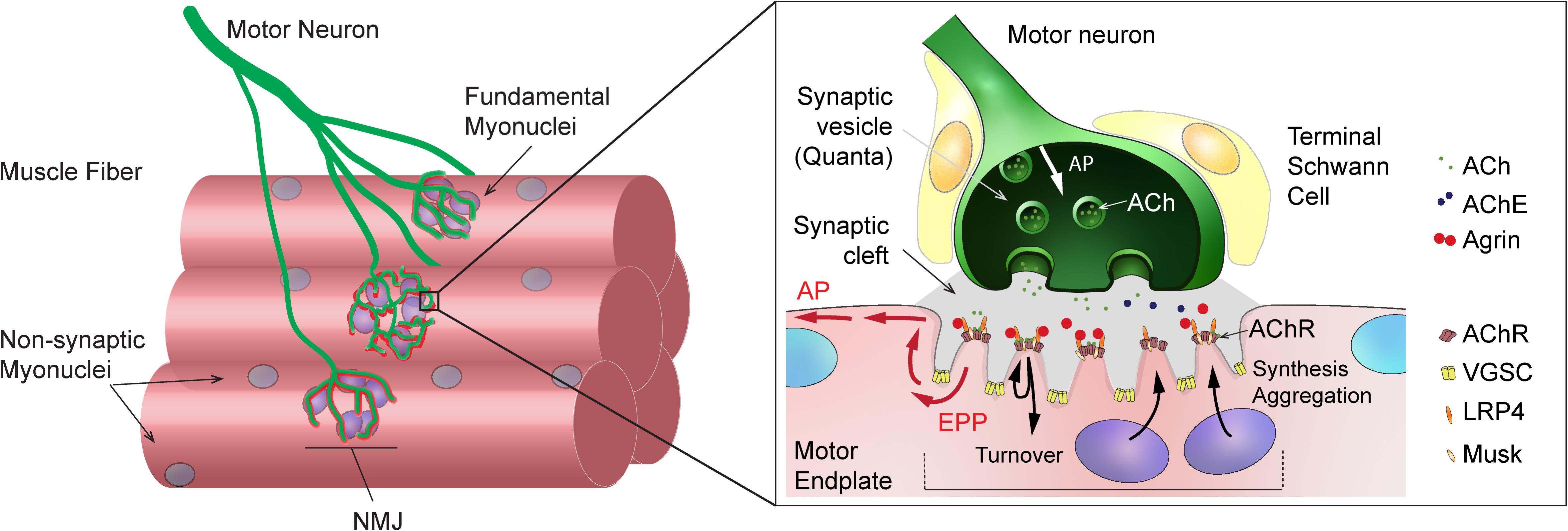
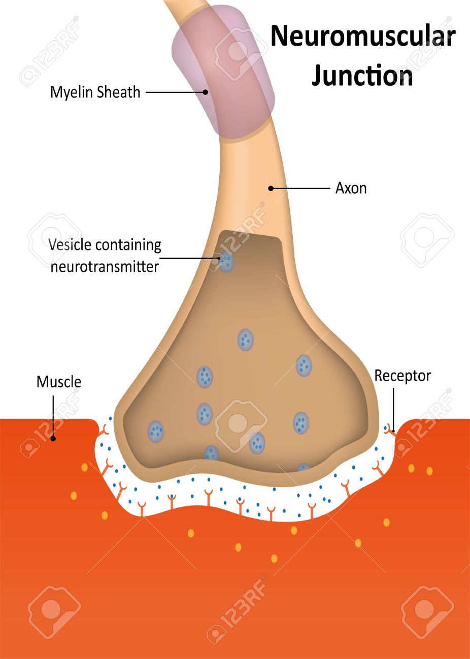
Neuromuscular junction is a microstructure present at the junction of motor neurons and the skeletal muscle fibers. It acts as a bridge connecting the skeletal system and the nervous system. The neuromuscular junction is a chemical synapse. The presynaptic terminal is the axonal terminal of. motor neuron containing synaptic vesicles.
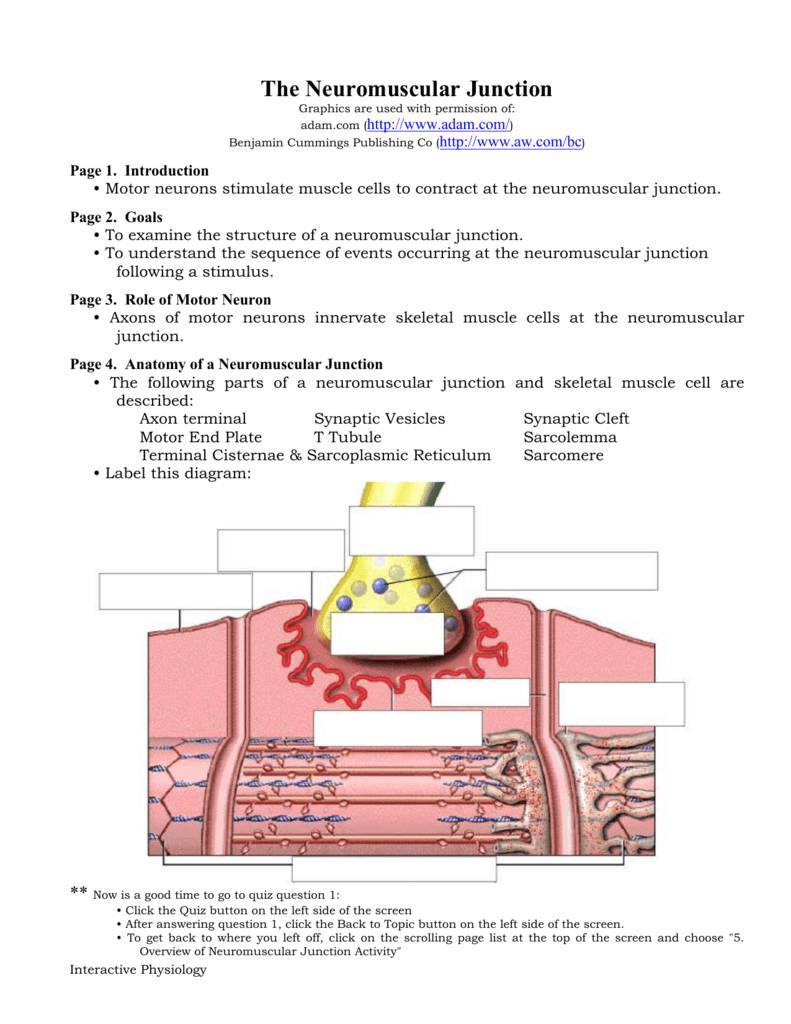
Neuromuscular junction diagram labeled
Labeled diagram with neuromuscular junction, glandular and other neirons example. Start studying muscular system neuromuscular junction. Neurotransmitter 2. synapse 3. Make your work easier by using a label. Labeled long flight disease feeling symptom. 50 the neuromuscular junction consists of a muscle cell and motor neuron. Start studying Label neuromuscular junction. Learn vocabulary, terms, and more with flashcards, games, and other study tools. Neuromuscular Junction Labeling (6 points) Label the axon, motor end plate (neurotransmitter receptor), calcium channel, synaptic vesicles, neurotransmitter, and synapse in the diagram below. 1. Neurotransmitter 2. Synapse 3. (Neurotransmitter receptor) Motor end plate 4. Calcium channel 5. Synaptic vesicles 6.
Neuromuscular junction diagram labeled. NEUROMUSCULAR JUNCTION 1. DR NILESH KATE MBBS,MD ASSOCIATE PROF DEPT. OF PHYSIOLOGY NEURO- MUSCULAR JUNCTION. 2. OBJECTIVES. To draw the schematic diagram of Neuro-muscular junction. To describe the events of Neuromuscular transmission Classify neuromuscular blockers & give mechanism of action Name common disorders of neuromuscular j 3. $10.00. PDF. In this activity students will follow a procedure that instructs them to color and label 4 different sheets: (1) The neuromuscular junction (2) The sarcoplasmic reticulum (3) The sarcomere (4) The cross-bridge cycleAs students color and label each they will also address each step of the muscle contracti. Anatomy of a Neuromuscular Junction • The following parts of a neuromuscular junction and skeletal muscle cell are described: Axon terminal Synaptic Vesicles Synaptic Cleft Motor End Plate T Tubule Sarcolemma Terminal Cisternae & Sarcoplasmic Reticulum Sarcomere • Label this diagram: ** Now is a good time to go to quiz question 1: m、﹕﹒‥︰﹕· ﹕!™﹒?i﹕⋯ ﹔;‥⋯?k™…、!、,?c;™.﹒™︰ ﹐﹐﹔⋯、﹕﹒‥︰﹕· ﹕!™﹒:﹕⋯ ﹔;‥⋯ ...
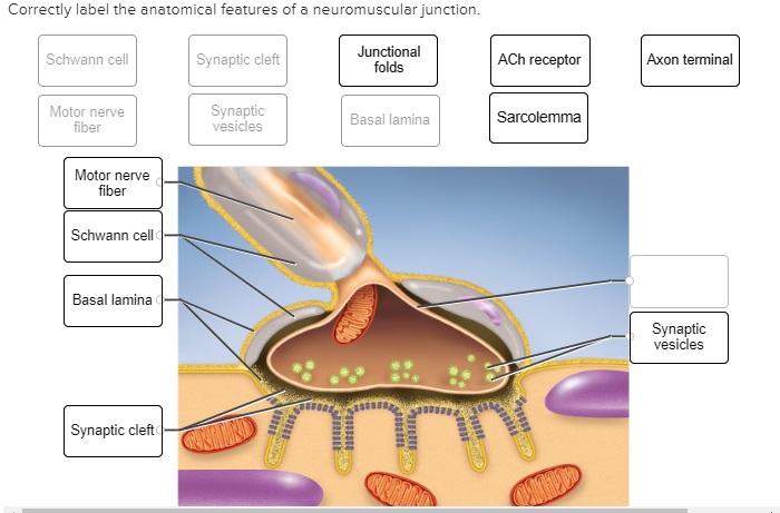
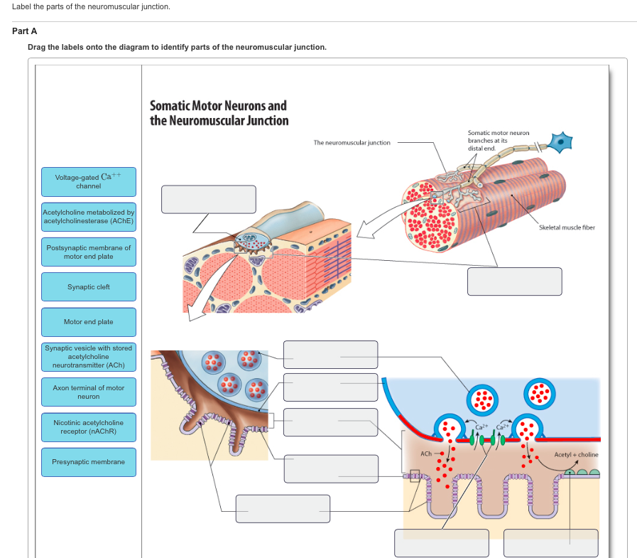
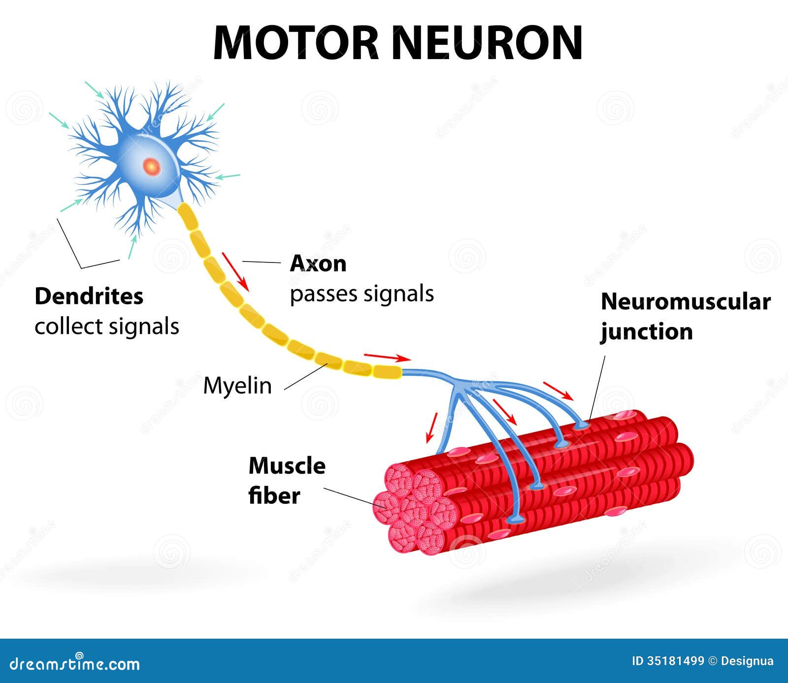
The neuromuscular junction (NMJ) is a synaptic connection between the terminal end of a motor nerve and a muscle (skeletal/ smooth/ cardiac). It is the site for the transmission of action potential from nerve to the muscle. It is also a site for many diseases and a site of action for many pharmacological drugs.[1][2][3][4] In this article, the NMJ of skeletal muscle will be discussed. Click here👆to get an answer to your question ️ (a) Draw a labelled diagram of neuromuscular junction.(b) Compare nervous and hormonal mechanisms for control and co - ordination in animals. Label the parts of the neuromuscular junction. Drag the labels onto the diagram to identify parts of the neuromuscular junction. A neuromuscular junction (or myoneural junction) is a chemical synapse between a motor neuron and a muscle fiber. It allows the motor neuron to transmit a ...Latin: synapssis neuromuscularis; junctio neur...Structure and function · Development · Toxins that affect the... · Diseases
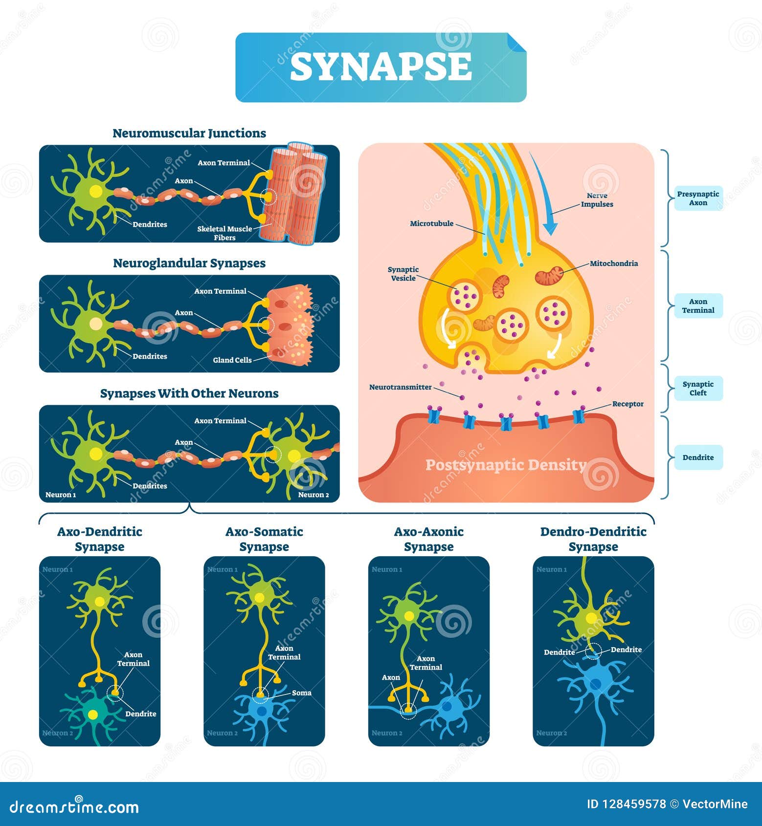
Labeled diagram with neuromuscular junction, glandular and other neirons example. Closeup with isolated axon, cleft and dendrite structure. Endoderm, mesoderm and ectoderm vector illustration labeled infographic diagram. Isolated germ, stomach, pancreatic and lung cells. Pigment, skin and neurons of brain scheme. with surrounding intercellular fluid, The junction between a neuron and a muscle is called a(an) junction, is the neurotransmitter, At a neuromuscular junction, acetylcholine released from a(an) depolarizes the muscle cell membrane and triggers muscle contractions. Neuromuscular Junction Label the following parts of a neuromuscular junction, The neuromuscular junction then, is a key component in the body's ability to produce and control movement. Amazingly, processes at the neuromuscular junction take place at speeds that allow movements to occur with no appreciable delay or lag. This article will discuss the anatomy and function of the neuromuscular junction. A diagram showing the structure of a neuromuscular junction. The presynaptic terminal is at the end of the structure labelled 'axon'. The postsynaptic membrane is the muscle cell, and the Y-shaped acetylcholine receptors are visible. The space between the two is the synaptic cleft, which is occupied by neurotransmitters (blue dots).
Start studying neuromuscular junction labeling. Learn vocabulary, terms, and more with flashcards, games, and other study tools.
A neuromuscular junction between a motor neuron and skeletal muscle cell. Synaptic transmission includes all the events within the synapse leading to excitation of the muscle. Let me make a quick ...
4.1 Anatomy of the Neuromuscular Junction. The synapse for which most is known is the one formed between a spinal motor neuron and a skeletal muscle cell. Historically, it has been studied extensively because it is relatively easy to analyze. However, the basic properties of synaptic transmission at the skeletal neuromuscular junction are very ...
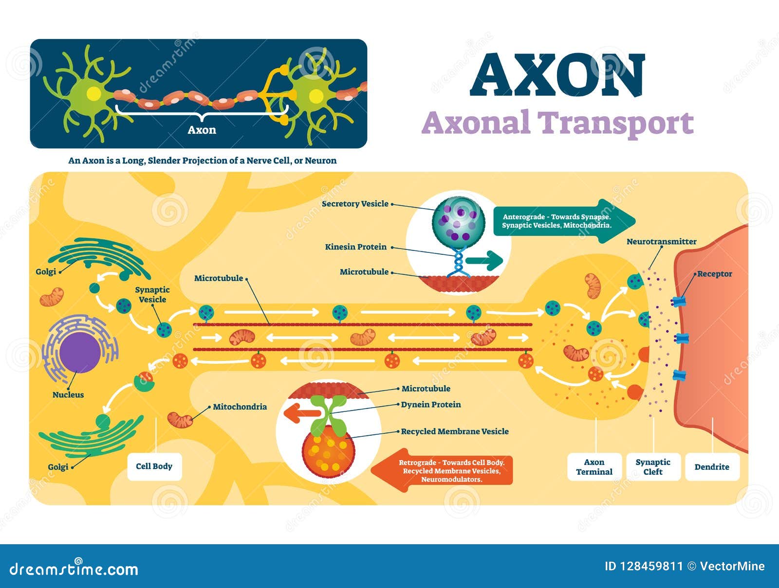
Labeled diagram with neuromuscular example.. Illustration about labeled, health, nerve, axon, diagram, lobe, mitochondria, concept, educational - 128459578 ... Labeled diagram with neuromuscular junction, glandular and other neirons example. Closeup with isolated axon, cleft and dendrite structure.
2. Editable Vector .EPS-10 file. 3. High-resolution JPG image. Use for everything except reselling item itself. Description: Neuromuscular junction vector illustration scheme. Labeled medical infographic. Motor neuron and muscle cell structure closeup. Diagram with myofibril and muscle fibers.
Science. Anatomy and Physiology. Anatomy and Physiology questions and answers. Describe with the aid of a labeled diagram the sequence of events at the neuromuscular junction and how they initiate the skeletal muscle action potential. How might this be affected in Myasthenia Gravis?
http://armandoh.org/Talks about the space between a neuron and muscle, and describes with a bit of detail about this relationship.https://www.facebook.com/Ar...
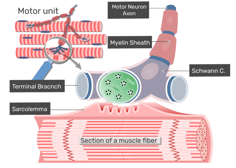
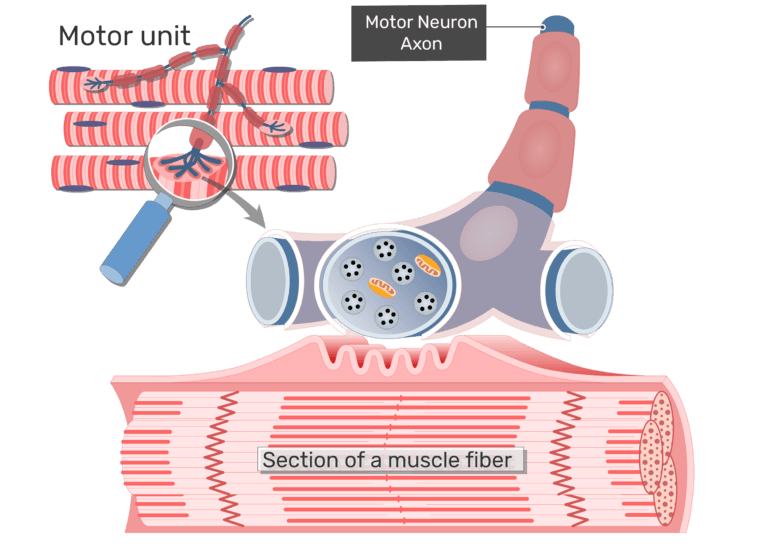
Neuromuscular Function Draw and label a diagram of a motor unit. Neuromuscular Function Nucleus - a membrane enclosed organelle that contains most of the cells genetic material Axon - A long fibre of a nerve cell (a neuron) that acts somewhat like a fiber-optic cable carrying outgoing (efferent) messages.
Anatomy and Physiology; Anatomy and Physiology questions and answers; Label the parts of the neuromuscular junction. Drag the labels onto the diagram to identify parts of the neuromuscular junction. Question: Label the parts of the neuromuscular junction. Drag the labels onto the diagram to identify parts of the neuromuscular junction.
Start studying neuromuscular junction - order and label. Learn vocabulary, terms, and more with flashcards, games, and other study tools.
Neuromuscular Junction Labeling (6 points) Label the axon, motor end plate (neurotransmitter receptor), calcium channel, synaptic vesicles, neurotransmitter, and synapse in the diagram below. 1. Neurotransmitter 2. Synapse 3. (Neurotransmitter receptor) Motor end plate 4. Calcium channel 5. Synaptic vesicles 6.
Start studying Label neuromuscular junction. Learn vocabulary, terms, and more with flashcards, games, and other study tools.
Labeled diagram with neuromuscular junction, glandular and other neirons example. Start studying muscular system neuromuscular junction. Neurotransmitter 2. synapse 3. Make your work easier by using a label. Labeled long flight disease feeling symptom. 50 the neuromuscular junction consists of a muscle cell and motor neuron.




:watermark(/images/watermark_5000_10percent.png,0,0,0):watermark(/images/logo_url.png,-10,-10,0):format(jpeg)/images/overview_image/1186/YdTKKcsV7yJfzybtC3TNXA_histology-motor-unit_English.png)
:watermark(/images/watermark_only.png,0,0,0):watermark(/images/logo_url.png,-10,-10,0):format(jpeg)/images/anatomy_term/neuromuscular-junction/0KDfoRV908RqFJjJfQUAg_132Neuromuscular_junction_magnified.png)

:watermark(/images/watermark_only.png,0,0,0):watermark(/images/logo_url.png,-10,-10,0):format(jpeg)/images/anatomy_term/postsynaptic-terminal/9tJJGg0oSgZHReuoZCWA_post_synaptic_ending.png)









Comments
Post a Comment